Understanding Pressure on the Optic Nerve
Intro
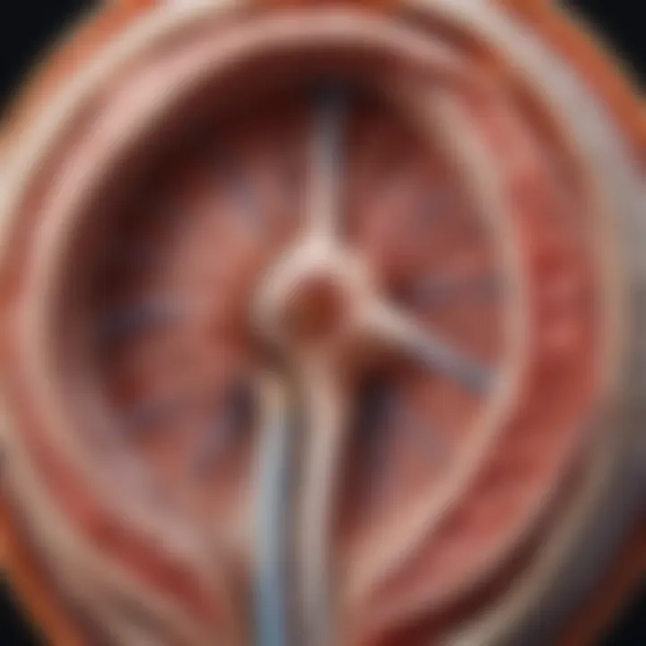
The optic nerve plays a crucial role in vision, transmitting visual information from the retina to the brain. However, the pressure exerted on this nerve can lead to significant health issues. Understanding the implications of increased pressure on the optic nerve is essential for maintaining eye health and preventing vision loss. This article aims to provide a comprehensive discussion on the anatomical structure of the optic nerve, causes of increased pressure, diagnostic methods, and treatment options, along with preventive strategies and future research areas.
Research Overview
Methodological Approaches
Research into optic nerve pressure has utilized various methodological approaches. Clinical studies often measure intraocular pressure (IOP) through tonometry, allowing for the assessment of potential risk factors associated with elevated pressure. Imaging techniques, such as optical coherence tomography (OCT), provide detailed views of the optic nerve's structure, enabling the identification of changes correlated with optic nerve pressure. Furthermore, animal models have been integral in understanding the biological mechanisms that underlie increased pressure and its consequences on ocular health.
Significance and Implications
Understanding the impact of pressure on the optic nerve goes beyond academic interest; it holds clinical significance. High intraocular pressure is a known risk factor for glaucoma, a leading cause of blindness.
According to the World Health Organization, glaucoma affects over 60 million people globally, underscoring the need for effective diagnosis and treatment strategies.
By comprehending the mechanisms of optic nerve pressure, healthcare professionals can enhance patient care, leading to timely interventions that may preserve vision.
Current Trends in Science
Innovative Techniques and Tools
The field of ophthalmology is witnessing rapid advancements. Innovative techniques, such as minimally invasive procedures and enhanced imaging modalities, are changing how practitioners diagnose and manage increased optic nerve pressure. Additionally, the development of telemedicine tools has made it easier for patients to receive evaluations remotely, improving access to care.
Interdisciplinary Connections
There is an increasing trend towards interdisciplinary research involving neurology, genetics, and even artificial intelligence. Collaborative efforts are yielding new insights into the pathophysiology of optic nerve pressure, potentially leading to the development of novel therapeutic targets. Involving multiple fields broadens the understanding and approach to managing this critical aspect of eye health.
Prologue to the Optic Nerve
The optic nerve is a critical structure in the visual system. It serves as the main pathway for transmitting visual information from the retina to the brain. Understanding its anatomy and function is key to appreciating the various kinds of disorders that can arise from pressure on the optic nerve. This pressure can affect sight and overall eye health significantly, leading to a range of complications if not addressed.
In this article, we will explore the intricacies of the optic nerve, examining its anatomy and the mechanisms through which it functions. We will delve into the clinical significance of pressure metrics on this nerve, highlighting why monitoring these values is essential for maintaining proper visual health. Whether you are a student, a researcher, or a professional in ocular health, a solid grasp of these foundational concepts will enhance your understanding of the implications of increased pressure on the optic nerve.
Anatomy of the Optic Nerve
The optic nerve is roughly cable-like in structure, composed of around one million nerve fibers. These fibers originate in the ganglion cells of the retina and converge to form the optic nerve head, or papilla. The nerve then travels through the optic canal in the skull. At this point, the optic nerves from both eyes partially cross at the optic chiasm. This crossing allows visual information from both fields to be processed together, enhancing depth perception and visual clarity.
Moreover, the diameter of the optic nerve ranges from 1.5 to 2.0 mm at the optic disc and tapers as it progresses towards the brain. The nerve is covered by three layers of protective sheaths. Understanding this anatomy is crucial for diagnosing various pathologies related to increased pressure, such as glaucoma or optic neuritis.
Function of the Optic Nerve
The function of the optic nerve is primarily to convey visual information. When light enters the eye, it is converted into electrical signals in the retina. These signals are then transmitted through the optic nerve to the brain, where they get processed into images. This transformation is essential for activities ranging from simple object recognition to complex tasks requiring spatial awareness and judgment.
Pressure on the optic nerve can disrupt this pathway. Elevated pressure can distort the nerve fibers, leading to a reduction in the clarity of visual signals. Symptoms may include blurred vision, loss of peripheral vision, or even complete loss of sight. Therefore, recognizing the functions of the optic nerve is paramount in comprehending the broader implications of conditions that lead to increased pressure.
What is Pressure on the Optic Nerve?
Understanding pressure on the optic nerve is crucial for grasping its implications on vision health. The optic nerve plays a vital role in transmitting visual information from the eye to the brain. When excessive pressure affects this nerve, it can lead to serious visual disturbances and other complications. The significance of this topic is underpinned by the interplay between the optic nerve's anatomical structure and its function. Establishing the correct context around optic nerve pressure not only aids in diagnosing but also in managing conditions related to it.
Definition and Clinical Significance
The term "optic nerve pressure" refers to the stress or tension exerted on the optic nerve, typically due to increased intracranial pressure. Clinically, this pressure can be measured and assessed through various diagnostic methods. Failure to address abnormal pressure levels can result in irreversible damage to the nerve, leading to vision loss. Conditions like glaucoma, tumors, and idiopathic intracranial hypertension are commonly associated with this phenomenon. These conditions highlight the importance of understanding optic nerve pressure, as timely intervention can significantly alter patient outcomes. To fully appreciate the clinical significance, one should consider the lifecycle of optic nerve health, which can be disrupted by prolonged elevated pressure.
Normal Pressure Ranges
Normal pressure ranges on the optic nerve are generally defined based on intraocular pressure (IOP) and intracranial pressure metrics. Typically, a healthy IOP is considered to be between 10 to 21 mmHg. However, normal intracranial pressure should be within 7-15 mmHg. It is crucial to understand that pressure readings outside these ranges may indicate potential disorders requiring medical attention. For instance, an IOP exceeding the normal range may suggest glaucoma, while an intracranial pressure higher than normal signifies potential neurological concerns.
Understanding the normal ranges of optic nerve pressure is essential for effective diagnosis and treatment planning.
Knowing the baseline values allows healthcare professionals to identify deviations that might signal a problem. An early diagnosis can lead to timely management strategies that can preserve vision and overall eye health. Regular monitoring of pressure values is therefore a necessary component of eye health management.
Causes of Increased Pressure
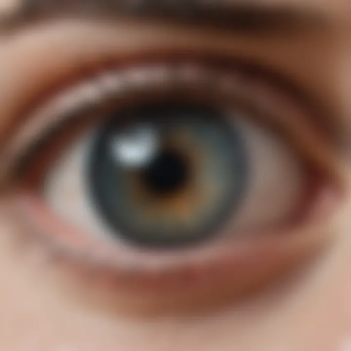
Understanding the causes of increased pressure on the optic nerve is essential for addressing potential vision loss and other complications. It allows for early diagnosis and intervention, which can have significant implications for maintaining visual health. Several conditions contribute to this increase in pressure, including glaucoma, tumors, optic neuritis, and idiopathic intracranial hypertension. Each of these factors can reveal critical insights into how pressure buildup occurs and what potential responses might be necessary.
Glaucoma
Glaucoma is one of the main causes of increased pressure on the optic nerve. This condition is characterized by a gradual loss of optic nerve fibers, often linked to elevated intraocular pressure (IOP). The primary concern with glaucoma is its stealthy nature; it can progress without noticeable symptoms until substantial damage has occurred.
The increased pressure in the eye can result from an imbalance in the production and drainage of aqueous humor. If the drainage channels become blocked or are overly constricted, pressure rises. The consequences can be devastating, leading to irreversible vision loss if not treated promptly. Treatments may include medications like prostaglandin analogs to enhance drainage or surgical options aimed at improving fluid outflow.
Tumors and Masses
Tumors and masses can also cause increased optic nerve pressure. Both benign and malignant growths can exert pressure on the optic nerve as they expand within the cranial cavity or orbits. The location and size of the tumor significantly impact its effects. For instance, a pituitary adenoma can push against the optic chiasm, leading to visual field defects.
Diagnosing these conditions may require imaging techniques such as magnetic resonance imaging (MRI) or computed tomography (CT). Management strategies often involve surgical removal of the tumor, radiation therapy, or chemotherapy, depending on the tumor’s nature and malignancy.
Optic Neuritis
Optic neuritis is an inflammatory condition that can lead to increased pressure on the optic nerve. In many cases, it is associated with multiple sclerosis, a disease that attacks the nervous system. Inflammation can cause swelling and a rise in pressure, producing symptoms like blurry vision or color distortion.
Diagnosis is often confirmed through an eye examination, MRI scans, or visual field tests. Treatment may involve corticosteroids to reduce inflammation and improve recovery, although the effectiveness can vary based on the individual.
Idiopathic Intracranial Hypertension
Idiopathic intracranial hypertension (IIH) is another significant cause of heightened optic nerve pressure. This condition occurs when there is increased pressure around the brain without an apparent cause. Symptoms may include headache, visual disturbances, or pulsatile tinnitus, making recognition vital.
Diagnosing IIH typically involves lumbar puncture to measure cerebrospinal fluid (CSF) pressure and evaluate the presence of other conditions. Management strategies often include lifestyle modifications, weight loss, diuretics, and in severe cases, surgical procedures to relieve pressure.
The awareness and understanding of these causes can empower healthcare professionals to intervene strategically and improve patient outcomes.
Symptoms Associated with Excess Pressure
Increased pressure on the optic nerve often manifests through various symptoms. Recognizing these symptoms is crucial for timely diagnosis and treatment. Such awareness can prevent serious complications, including vision loss. Symptoms can vary significantly, which demands a tailored approach for evaluation and management. The common symptoms include visual disturbances, headaches, and nausea or vomiting. Each will be discussed to provide a clear understanding of their significance.
Visual Disturbances
Visual disturbances can present as blurriness, blind spots, or even the perception of flashing lights. These disturbances often signal that the optic nerve is under duress. When pressure increases, it may compress the nerve fibers responsible for vision. As a result, one may experience temporary or persistent changes in sight.
Understanding the mechanisms that lead to these disturbances is vital. Increased pressure might lead to reduced blood flow, thereby compromising the optic nerve's function. Early recognition of these symptoms is key. If left unchecked, these disturbances can escalate, underpinning the importance of seeking medical attention promptly.
Headaches
Headaches represent another common symptom linked to excess pressure on the optic nerve. They can range from mild discomfort to debilitating pain. Often, individuals may describe these headaches as pulsating or throbbing, which may coincide with visual changes. The connection between headache and optic nerve pressure arises from the brain's anatomical proximity to the optic nerve. Increased intracranial pressure can stimulate pain receptors, resulting in headache.
Evaluating headache patterns is essential for health professionals. Those experiencing persistent or unusual headaches should consider the context of other symptoms. Accurate assessment can lead to essential interventions before more severe outcomes ensue.
Nausea and Vomiting
Nausea and vomiting are less frequently discussed symptoms but remain significant indicators of high optic nerve pressure. These symptoms may result from the brain's reaction to elevated pressure. Increased pressure can affect the centers in the brain that regulate nausea. When these symptoms occur alongside visual disturbances or headaches, they merit further investigation.
Taking these symptoms seriously is necessary for diagnosis and treatment. Many may overlook nausea, attributing it to unrelated factors. However, when present with other visual or headache symptoms, it could signal optic nerve pressure. Immediate medical consultation is advised to rule out underlying conditions.
Diagnosis of Optic Nerve Pressure Conditions
Diagnosis of conditions affecting the optic nerve pressure is crucial for maintaining visual health. Identifying increased pressure early can help prevent serious complications. Understanding these diagnostic methods allows clinicians to tailor interventions and optimize patient outcomes. Notably, the relationship between symptoms and the underlying pressure is complex. Thus, tools that provide a clearer picture are vital.
Effective diagnosis contributes not only to treatment planning but also to educating patients about the conditions affecting their vision. It paves the path for ongoing monitoring and management strategies. The primary aim remains to preserve visual function and improve quality of life.
Optical Coherence Tomography
Optical Coherence Tomography (OCT) serves as a non-invasive imaging technique. It provides cross-sectional images of the optic nerve head and retina. By measuring the thickness of the retinal nerve fiber layer, this method can indicate potential damage from increased pressure. OCT enables early detection of optic nerve issues before significant visual impairment occurs.
Some advantages of OCT include:
- High resolution: Allows for detailed views of ocular structures.
- Non-contact nature: Minimizes patient discomfort.
- Fast imaging: Reduces time spent in the clinic.


Regular OCT assessments can help track changes over time, assisting in the management of conditions like glaucoma.
Visual Field Testing
Visual field testing remains a cornerstone in optic nerve pressure evaluation. It assesses peripheral vision, which can be affected by increased pressure. By identifying blind spots or changes in vision, clinicians can ascertain if the optic nerve is being compromised.
Two common approaches in visual field testing are:
- Static perimetry: Involves detecting light spots on a blank background.
- Kinetic perimetry: Measures the point at which a moving light stimulus is perceived.
Both methods provide essential data regarding visual function. Timely visual field tests can indicate disease progression, leading to adjustments in treatment plans.
Fundoscopy Examination
Fundoscopy examination involves direct observation of the optic nerve head through the pupil. This technique helps identify swelling or changes in color that suggest increased pressure. Fundoscopy is essential for assessing the health of the optic nerve and the retina.
Some key aspects of a fundoscopy include:
- Optic disc evaluation: Observing the margins and the cup-to-disc ratio.
- Retinal examination: Checking for any signs of hemorrhage or edema.
- Direct insights: Provides immediate feedback on nerve health.
Both the structural status of the optic nerve and changes related to systemic conditions can be monitored through a thorough fundoscopy. This examination often leads to further investigations or immediate interventions if abnormalities are detected.
The accuracy of these diagnostic tools is paramount in mitigating the risks associated with optic nerve pressure. Timely and precise detection can significantly impact treatment options and outcomes.
Treatment Options for High Optic Nerve Pressure
Understanding the treatment options for high optic nerve pressure is crucial. These treatments aim not only to alleviate pressure but also to prevent potential damage to the optic nerve, which can lead to vision impairment. Each treatment type has its own benefits and specific considerations. Managing increased pressure effectively can improve both immediate and long-term visual outcomes for patients.
Medications
Medications are often the first line of treatment when addressing high optic nerve pressure. The primary goal is to reduce fluid accumulation or minimize factors contributing to increased intracranial pressure. Common medications include:
- Carbonic Anhydrase Inhibitors: These reduce the production of cerebrospinal fluid, thus lowering pressure. Acetazolamide is a well-known drug in this class.
- Diuretics: Medications like furosemide can help the body eliminate excess fluid.
- Corticosteroids: These are used to reduce inflammation that may contribute to optic nerve swelling.
It's important for patients to adhere to prescribed regimens and discuss any side effects with healthcare providers. Additionally, monitoring the efficacy of medications can determine if adjustments are needed to optimize treatment.
Surgical Interventions
In cases where medications are insufficient or not tolerated, surgical interventions might be necessary. Surgical options can provide significant relief from elevated optic nerve pressure. The following procedures are commonly performed:
- Optic Nerve Decompression: This surgery entails removing bone or tissue that is compressing the optic nerve, allowing for increased space and reduced pressure.
- Shunt Placement: In certain situations, a shunt can be implanted to facilitate the drainage of excess cerebrospinal fluid, thereby lowering pressure.
Patients undergoing surgical interventions should discuss potential risks and complications with their healthcare provider. Individual assessments will help determine the most appropriate approach to ensure successful outcomes.
Monitoring and Follow-Up Care
Continuous monitoring and follow-up care are fundamental components of managing optic nerve pressure. Regular appointments allow for:
- Assessment of Treatment Efficacy: Tracking changes in pressure and visual acuity is vital. This includes reviewing any adjustments to medications or modifications in surgical plans.
- Detection of Complications: Continuous care offers the opportunity to identify any potential side effects or complications early on.
- Patient Education: Ongoing communication with healthcare teams ensures patients understand their condition and the importance of adherence to treatment strategies.
"Regular follow-up care is essential. It helps in managing both expectations and outcomes effectively."
Patients should not hesitate to bring up their concerns during follow-up visits. This open dialogue ensures better tailored treatment and helps in adapting strategies as needed.
Impact of Increased Pressure on Visual Health
Increased pressure on the optic nerve is a critical issue that can lead to significant visual health complications. Understanding this impact not only highlights the physiological ramifications but also initiates a discussion around prevention and management strategies. The optic nerve carries visual information from the eye to the brain, and any pressure on this nerve can disrupt the transmission of these signals. This can manifest in various ways, complicating patients' lives and limiting their ability to engage in everyday activities.
Potential for Vision Loss
The potential for vision loss due to increased pressure is profound. When the pressure rises beyond normal levels, the blood vessels in and around the optic nerve can be compromised. This deprivation hampers the oxygen and nutrients necessary for healthy nerve function, leading to irreversible damage. Conditions such as glaucoma are well documented for causing gradual vision loss as a result of chronic pressure buildup.
Moreover, studies show that patients with untreated high optic nerve pressure frequently experience visual field defects. Specific symptoms may include blurred vision, tunnel vision, and even complete blindness in advanced stages if not properly managed. The severity of vision loss often correlates with the duration and magnitude of the pressure increase. Thus, early detection and treatment are paramount in reducing the risk of permanent sight impairment.

Long-Term Consequences
The long-term consequences of increased pressure on the optic nerve extend beyond immediate vision issues. Chronic pressure conditions can change the anatomy of the optic nerve over time, leading to conditions like optic atrophy. This is characterized by the degeneration of the optic nerve fibers, which can result in profound and lasting vision deficits.
Furthermore, these changes can also lead to secondary complications, including psychological impacts such as anxiety and depression resulting from the loss of independence in daily activities. Patients may find it challenging to navigate their environments, leading to a diminished quality of life.
In summary, increased pressure on the optic nerve presents serious risks for both acute and chronic visual health implications. Early diagnosis and intervention are essential to mitigate the potential for vision loss, emphasizing the need for focused preventive measures and ongoing research on optic nerve health.
"Addressing increased optic nerve pressure through education and awareness can significantly impact patient outcomes."
Regular eye examinations and lifestyle modifications can serve as pivotal preventative strategies, ensuring patients remain vigilant about their eye health. Lack of attention to these issues may lead to irreversible damage, thus highlighting the necessity of both awareness and proactive management.
Preventative Measures
Preventative measures play a crucial role in maintaining optic nerve health and overall visual function. The implications of increased pressure on the optic nerve can lead to severe, sometimes irreversible, vision loss. Therefore, understanding and implementing effective strategies to reduce risk factors is paramount. In this section, we will examine lifestyle modifications and the importance of regular eye examinations.
Lifestyle Modifications
Adopting specific lifestyle changes can significantly mitigate the risk of developing conditions that cause increased optic nerve pressure. Consider the following lifestyle modifications:
- Healthy Diet: A balanced diet rich in antioxidants supports eye health. Nutrients like vitamins C and E, zinc, and omega-3 fatty acids can contribute to maintaining optimal vision. Including leafy greens, fruits, nuts, and fatty fish in your meals can provide these benefits.
- Regular Exercise: Engaging in physical activity helps maintain a healthy weight and can lower the risk of developing conditions such as diabetes and hypertension, which put extra strain on the optic nerve. Aim for at least 150 minutes of moderate aerobic activity each week.
- Stress Management: Chronic stress can contribute to various health issues, including those affecting the eyes. Techniques such as meditation, yoga, or deep breathing can assist in stress reduction.
- Avoid Tobacco and Excess Alcohol: Smoking and excessive alcohol consumption are linked to a higher risk of eye diseases. Eliminating these habits can help preserve vision and reduce optic nerve strain.
Incorporating these lifestyle choices into daily routines not only protects against optic nerve pressure but also boosts overall health.
Regular Eye Examinations
Regular eye examinations are vital for early detection and management of conditions linked to increased pressure on the optic nerve. These exams can identify issues before they result in significant damage. Key reasons to prioritize eye exams include:
- Baseline Assessment: Establishing a baseline visual assessment allows for effective monitoring of any changes over time. It provides a reference point for future examinations.
- Monitoring Eye Pressure: Eye care professionals typically check intraocular pressure during these examinations. If pressure is found to be elevated, further investigation and intervention can be initiated promptly.
- Early Detection of Disorders: Regular check-ups can help in identifying diseases like glaucoma or diabetic retinopathy in their early stages, allowing for timely treatment.
- Personalized Care: Continuous eye health monitoring enables doctors to tailor recommendations based on individual risk factors such as family history or pre-existing conditions.
End
In summary, preventative measures including lifestyle modifications and regular eye examinations are essential in combating increased pressure on the optic nerve. By taking action now, individuals can protect their visual health, reduce risk factors, and potentially avert significant vision loss down the line. Awareness and proactivity in eye care can lead to improved life quality while safeguarding one of our most precious senses.
Future Research Directions
Research into optic nerve pressure is crucial for enhancing our understanding and management of related conditions. Continuing investigations in this area have the potential to uncover new insights that influence both diagnostics and treatment strategies. The focus on research is not only about advancing medical knowledge; it also addresses real-world implications for patients experiencing vision-related issues.
Innovations in Diagnostics
Emerging technologies are significantly changing how we approach the diagnosis of optic nerve pressure conditions. For instance, machine learning algorithms are increasingly being utilized to analyze complex data from optical coherence tomography. This technology allows for detailed imaging of the optic nerve head and surrounding tissues, offering a more comprehensive view than traditional methods. The integration of artificial intelligence can enhance the accuracy of diagnosing subtle changes in optic nerve appearance, which may indicate early signs of increased pressure.
Furthermore, research into biomarkers for optic nerve pressure is of particular interest. Identifying specific molecular markers could enable blood tests that detect changes linked to elevated pressure. This non-invasive method may revolutionize how we monitor at-risk individuals.
Novel Therapeutic Approaches
The exploration of novel therapeutic approaches is vital for developing effective treatments for conditions characterized by increased optic nerve pressure. Current medications primarily aim to lower intraocular pressure; however, innovative strategies are on the horizon. For instance, gene therapy is being examined as a way to address the underlying causes of optic nerve damage. By targeting specific genes involved in ocular health, there is potential to restore function or protect the optic nerve from further deterioration.
Another area of interest is the development of neuroprotective agents. These compounds focus on preserving the health of the optic nerve amid increased pressure. Research shows promise in agents that minimize oxidative stress and inflammation, two critical factors in optic nerve damage.
Additionally, minimally invasive surgical techniques for managing optic nerve pressure are under investigation. These techniques aim to reduce complications and recovery time while effectively addressing the underlying issues.
"Investing in research on optic nerve pressure not only informs clinical practices but also uplifts the quality of care for patients facing vision issues."
Epilogue
Understanding the implications of pressure on the optic nerve is crucial for maintaining visual health. Increased pressure can lead to significant consequences, making it vital for individuals to recognize the risks associated with this condition. This article underscores the importance of identifying the causes of pressure, the symptoms that may arise, and the diagnostic tools available to evaluate the condition effectively.
Summary of Key Findings
In summary, pressure on the optic nerve can stem from various factors, including glaucoma, tumors, and idiopathic intracranial hypertension. The clinical significance of this pressure cannot be understated, as it often leads to visual disturbances and, if left untreated, could result in permanent vision loss.
Effective diagnosis requires advanced techniques such as Optical Coherence Tomography and thorough visual field testing. Treatment options range from medications to surgical interventions, contributing to the management of the condition. Monitoring and regular follow-up care remain essential in mitigating risks associated with prolonged elevated pressure.
Importance of Awareness and Education
Raising awareness about pressure on the optic nerve plays a pivotal role in preventing severe outcomes. Educating the public, along with professionals in the field, can lead to early detection and timely intervention. Awareness initiatives should focus on the symptoms of increased pressure, the significance of regular eye examinations, and the lifestyle modifications that can help reduce the risk.
Ultimately, understanding the mechanisms behind optic nerve pressure not only aids in clinical settings but also empowers individuals to take charge of their eye health. Keeping informed will allow for better health outcomes and contribute towards reducing the burden associated with vision-related disorders.


