Understanding Lung Nodules Detected on X-Ray
Intro
Lung nodules are small masses of tissue found in the lungs and are often identified through routine X-ray examinations. Their presence can raise significant concerns, making it imperative to understand their nature. These nodules can vary in size and appearance. In many cases, they are benign and pose little to no threat. However, determining whether a nodule is benign or malignant is critical for patient management.
When a lung nodule is detected, it often leads to further diagnostic procedures. For instance, the characteristics of the nodule, such as its size, shape, and edge smoothness, can provide clues regarding its nature. Some nodules will require additional imaging techniques like CT scans to give a clearer picture, while others may necessitate more invasive procedures, including biopsies.
This article will delve into the complexities surrounding lung nodules, providing an in-depth look at various aspects, including diagnostic processes and clinical management. By exploring key differentiators between benign and malignant nodules and discussing contemporary techniques associated with their diagnosis and treatment, we aim to clarify this crucial aspect of pulmonary health.
Preface to Lung Nodules
Lung nodules are small masses within the lung that can raise significant concerns regarding pulmonary health. Detecting these nodules during routine examinations is crucial, as they may signal serious underlying issues. This section establishes the foundation for understanding lung nodules, their implications, and their relevance in medical diagnostics.
The importance of this topic cannot be overstated. Lung nodules can vary greatly in nature; some are benign, while others may represent malignancy. By defining these nodules clearly, we can better assess their characteristics and potential risks. Awareness of lung nodules promotes timely diagnosis and potential treatment, which is critical in improving patient outcomes.
Moreover, comprehending the prevalence and significance of lung nodules helps the medical community address pulmonary conditions more effectively. Understanding the trends and risks associated with lung nodules guides both research and clinical practices, ultimately leading to improved patient management protocols.
Role of X-Ray in Lung Nodule Detection
Lung nodules can be detected during routine X-ray examinations, making the understanding of the role of X-ray crucial. X-rays serve as a first line of investigation to identify potential abnormalities in the lungs. They provide a non-invasive method for initial assessments. Their importance stems not only from their widespread availability but also from their ability to offer a quick overview of lung health. Not all nodules are dangerous, but identifying them is vital to determine necessary follow-up procedures.
Mechanism of X-Ray Imaging
X-ray imaging works by passing a controlled amount of radiation through the body. The dense structures, like bones, absorb more radiation, while less dense tissues, such as those in the lungs, allow more radiation to pass through. This differential absorption creates an image. The radiation that comes out of the body hits a film or sensor on the opposite side, forming an image depicting the internal structures. When it comes to lung nodules, X-ray imaging reveals shadows that may indicate the presence of a nodule.
X-rays are particularly effective for catching nodules that are larger than 3 millimeters. However, smaller nodules might not be detected. Thus, while X-rays offer a good starting point, their limitations necessitate further testing in certain cases.
Interpreting X-Ray Results
Interpreting X-ray results requires a trained eye. Radiologists assess various factors in the lung images. The shape, edges, and density of the identified nodules are essential in determining their nature. A smooth border might suggest a benign growth, while irregular or jagged edges could indicate malignancy. Additionally, the size of the nodule plays a key role. Nodules smaller than 6 millimeters are often monitored over time, while larger or suspicious nodules may require further diagnostic procedures such as CT scans or biopsies.
"X-ray interpretation is both an art and a science. It involves recognizing patterns and understanding their implications in the context of patient history and other tests."
Ultimately, X-ray results contribute significantly to the larger picture of lung health but must be interpreted cautiously. A multidisciplinary approach is necessary for accurate diagnosis and management of lung nodules.
Characteristics of Lung Nodules
Understanding the characteristics of lung nodules is vital in assessing their implications for pulmonary health. These nodules may indicate various issues ranging from benign anomalies to potential malignancies. Several specific elements contribute to the importance of this topic, notably size, shape, and radiological appearance. Evaluating these characteristics helps clinicians to make informed decisions regarding further testing and treatment options.
Size and Shape Variations
The size and shape variations of lung nodules are crucial factors in distinguishing between benign and malignant types. Nodules can be classified into small, medium, and large, with each category offering different insights. Small nodules, typically under 1 centimeter, are often benign but can necessitate follow-ups.
Medium nodules, ranging from 1 to 3 centimeters, require closer observation as they may present a higher risk for malignancy. Large nodules, exceeding 3 centimeters, are often treated with a more aggressive diagnostic approach due to the increased likelihood of cancer.
Shape also plays a pivotal role; round, well-defined nodules often suggest benign conditions, while irregular shapes with spiculated edges may indicate malignancy. These variations highlight the necessity for accurate measurement and description during diagnostic imaging.
Radiological Appearance
The radiological appearance of lung nodules provides new dimensions for analysis. Nodules can present in various forms, such as solid, subsolid, or ground-glass opacity. Solid nodules appear as dense white areas on X-rays and may require immediate investigation.
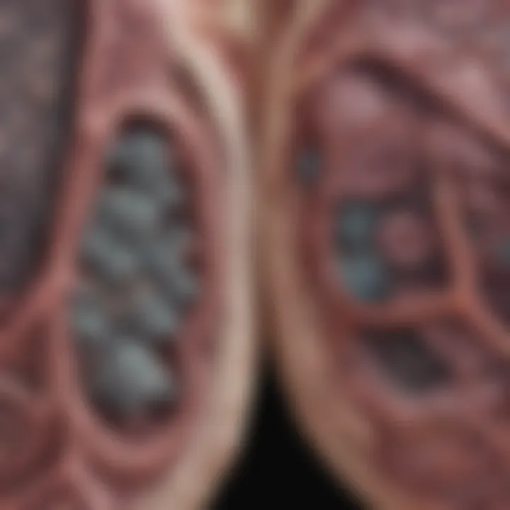
Subsolid nodules consist of both solid and ground-glass components; their characteristics can shift over time, raising concerns about their potential malignancy. Ground-glass opacities are often associated with a lower risk of cancer, but follow-up may still be required if changes are noted.
Understanding these characteristics enables healthcare providers to stratify risk and facilitate timely interventions when necessary.
Benign Versus Malignant Nodules
The distinction between benign and malignant lung nodules is critical in the evaluation process following their detection. This section explores the significance of understanding these two categories, implications for patient management, and the strategies for diagnostic differentiation.
Defining Benign Nodules
Benign nodules are typically non-cancerous growths that do not pose a significant health threat. They might result from infections, inflammatory diseases, or previous injuries in the lung. Common examples are hamartomas or granulomas.
Some key characteristics of benign nodules include:
- Size: Often smaller than malignant nodules.
- Growth Rate: Generally exhibit slow progression or remain stable over time.
- Appearance: Smooth edges and well-defined margins on imaging.
Knowing these traits helps healthcare providers approach treatment pathways effectively. While benign nodules usually require monitoring rather than aggressive intervention, patients benefit from clear communication regarding their benign nature.
Malignant Nodules: Risk Factors
In contrast, malignant nodules can indicate lung cancer or metastasis from other cancers. Understanding risk factors allows for better early detection and potentially more favorable outcomes. Some of the recognized risk factors for malignant nodules include:
- Smoking History: Individuals with a history of smoking are at a higher risk of developing lung cancer.
- Exposure to Carcinogens: Factors such as radon gas, asbestos, or industrial pollutants contribute to increased risk.
- Family History: A genetic predisposition may play a role in the development of lung malignancies.
- Age: Older individuals are more likely to experience malignant nodules compared to younger populations.
The identification of these risks not only aids in diagnosis but shapes surveillance strategies and management plans for at-risk patients, ensuring early intervention where needed.
Evaluating lung nodules as either benign or malignant is essential. This analysis informs further diagnostic testing and potential treatment protocols, which ultimately guides patient care. Understanding the differences improves patient outcomes by enabling timely and appropriate interventions.
Further Diagnostic Approaches
Lung nodules detected on X-rays can prompt further investigation to determine their nature. The initial findings can be inconclusive, necessitating additional techniques. This section explores the importance of follow-up diagnostics, including CT scans, PET scans, and biopsy procedures. Each approach offers unique insights that help differentiate benign from malignant nodules. Understanding these methods is crucial for effective patient management.
CT Scans: An Enhanced Imaging Technique
CT scans provide a more detailed view of lung nodules compared to standard X-rays. They use advanced imaging technology to produce cross-sectional images of the lungs. This allows for clearer visualization of the size, shape, and location of a nodule.
Benefits of CT scans include:
- Higher sensitivity: CT scans can detect smaller nodules that X-rays might miss.
- Characterization of nodules: Radiologists can analyze the internal structure of nodules, helping to evaluate risk factors.
- Monitoring changes: Serial CT scans can track the growth or changes in nodules over time, providing valuable data for clinical decisions.
While CT scans are highly effective, they do expose patients to higher levels of radiation than standard X-rays. Thus, the risks must be weighed against the benefits.
PET Scans for Metabolic Activity Assessment
PET scans assess not only the structure of lung nodules but also their metabolic activity. This technique measures the uptake of a radioactive glucose tracer, revealing how actively tissues are growing. Malignant tumors typically have a higher metabolic rate than benign ones.
Key points about PET scans:
- Dynamic analysis: PET scans can differentiate between active cancerous nodules and non-cancerous ones based on metabolic activity.
- Comprehensive view: They can also help identify additional malignancies, which might not be apparent during X-ray or CT imaging alone.
- Useful adjunct to CT: Combining PET and CT scans provides detailed anatomical and functional information, leading to more accurate diagnoses.

However, these scans are more expensive than traditional imaging. They also involve exposure to a small amount of radiation from the tracer. The decision to proceed with a PET scan should be based on clinical necessity.
Biopsies: When and Why
Biopsies are occasionally required when imaging results are inconclusive. A biopsy involves taking a small sample of tissue from the lung nodule for further examination. This procedure can confirm whether a nodule is benign or malignant and guide treatment decisions.
When to consider a biopsy:
- Suspicion of malignancy: If imaging indicates a high likelihood of cancer, a biopsy may be warranted.
- Growth or change in nodule appearance: Rapid changes in size or characteristics suggest the need for tissue analysis.
- Patient history: Previous cancer or significant risk factors could necessitate a biopsy, even for small nodules.
There are several types of biopsies, including:
- Needle biopsy: A minimally invasive option performed with imaging guidance.
- Surgical biopsy: More invasive but provides definitive results.
- Bronchoscopy: Allows for direct visualization and sampling of lung tissues.
Clinical Management of Lung Nodules
The clinical management of lung nodules is a crucial element in ensuring patients receive appropriate care. Effective management can influence outcomes significantly, making it imperative for healthcare providers to understand current protocols. With advancements in imaging technology and research, clinicians are better equipped to assess and manage lung nodules appropriately.
Surveillance Protocols
Surveillance protocols are designed to monitor lung nodules over time. This is especially important for nodules that show benign characteristics. Regular follow-ups can help in determining if a nodule changes in size or appearance.
Key considerations in surveillance include:
- Initial Assessment: After initial detection, the size and characteristics of the nodule are evaluated. For example, nodules less than 6 mm are often monitored for a longer duration, as they have a low risk of malignancy.
- Follow-Up Imaging: CT scans are usually employed for follow-ups, as they provide clearer images than X-rays. Typically, a follow-up protocol may include imaging every 3-12 months for the first couple of years, depending on the nodule's characteristics.
- Risk Stratification: Clinicians may categorize nodules based on risk factors such as smoker status or family history. Risk assessment can guide the frequency of surveillance needed.
"Regular follow-up is essential for effective management of lung nodules, balancing the needs for vigilance with reducing patient anxiety."
Treatment Options for Malignant Nodules
When lung nodules are determined to be malignant, treatment options must be discussed thoroughly with the patient. The approach taken often depends on the nodule's size, location, and overall health status of the patient.
Common treatment strategies include:
- Surgery: Surgical resection is often the preferred treatment for localized malignant lung nodules. Operations such as lobectomy or wedge resection may be performed to remove cancerous tissue.
- Radiation Therapy: For patients who are not surgical candidates or for those with advanced disease, radiation therapy can be an effective alternative.
- Chemotherapy: This is typically used in conjunction with other modalities for more advanced stages of lung cancer. It can help in managing symptoms and can be curative in some cases.
- Targeted Therapy: For certain types of lung cancer, targeted therapies can be employed. These medications aim at specific genetic markers or pathways related to the tumor's growth.
- Immunotherapy: An emerging treatment strategy that helps the immune system recognize and attack cancer cells more effectively.
In all cases, treatment plans should be individualized, taking into account patients' preferences, medical histories, and the unique characteristics of the nodules.
Patient Education and Support
Patient education and support is vital in the context of lung nodules detected on X-ray. Patients often experience anxiety and confusion upon hearing about any abnormalities in their lungs. Providing clear and accurate information can help alleviate fears and enable informed decisions about their healthcare.
Effective patient education begins with understanding the diagnosis. Patients should be informed about what lung nodules are, the likelihood of these nodules being benign or malignant, and the implications of their presence. Educators should focus on simplifying technical jargon while maintaining accuracy. This clarity aids patients in grasping the nature of their condition and enables them to engage in discussions with their healthcare providers. It's essential to emphasize that many lung nodules are harmless. Data suggests that over 90% of these nodules are benign, often resulting from infections, inflammation, or old scars. Awareness of these statistics can significantly reduce patient anxiety.
Understanding the Diagnosis
When patients receive a diagnosis of lung nodules, they might feel overwhelmed. It is crucial to explain the process of diagnosis step-by-step.
- Initial Detection: Nodules are often incidentally found on X-rays during unrelated examinations.
- Follow-up Imaging: Further imaging tests like CT scans are typically recommended for better evaluation.
- Assessment Process: With the help of radiologists, the characteristics of the nodule are assessed—its size, shape, and appearance play roles in determining the follow-up actions.
- Biopsy Consideration: For nodules that show concerning features, a biopsy may be necessary to determine if cancer is present.


Patients should be encouraged to ask questions about anything they do not understand. Having a clear explanation of their situation empowers patients, enabling them to make informed choices about their monitoring or treatment options.
Resources for Patients
Access to reliable resources can enhance patient education significantly. Many organizations and platforms provide comprehensive information regarding lung nodules and their management. Some notable resources include:
- American Lung Association: Offers detailed guides and FAQs on lung nodules.
- Mayo Clinic: Provides articles related to lung health and nodule-related information.
- WebMD: Contains accessible articles discussing the implications of lung nodules.
In addition, it is important for patients to build a support system. Local and online support groups can provide emotional comfort. Connecting with others who have had similar experiences offers valuable insights and reassurance.
Research shows that patients who actively engage in their healthcare are more likely to experience improved outcomes and satisfaction with their treatments.
Emerging Research and Future Directions
The landscape of lung nodule detection and management is undergoing rapid change, driven by advances in technology and research. As we enhance our understanding of lung nodules, it becomes critical to explore emerging research and future directions in this field. This section will cover novel imaging technologies and advancements in treatment modalities. Both aspects are essential for improving patient outcomes and optimizing clinical practices.
Novel Imaging Technologies
Recent years have witnessed significant progress in imaging technologies aimed at identifying and characterizing lung nodules. Traditional X-ray imaging has been a cornerstone of lung assessment, but limitations exist, notably concerning sensitivity and resolution.
Advancements such as 3D computed tomography (CT) scans and ultrasound techniques are now becoming more prevalent. These methods offer improved visualization of nodules, allowing for better characterization of size, shape, and density. Moreover, magnetic resonance imaging (MRI) is emerging as an alternative to CT scans in certain cases, particularly for patients who require multiple imaging sessions or are at risk for radiation exposure.
One notable development within imaging technology is machine learning and artificial intelligence. Implementing AI algorithms in radiology can enhance the detection rate of lung nodules on scans. Such technologies analyze vast amounts of data and learn from imaging patterns, potentially reducing human error. Notably, systems are now being designed to assist radiologists in prioritizing cases based on risk, leading to improved efficiency.
To fully realize these advancements, collaborative efforts among researchers, medical schools, and technology firms are vital. Studies are being conducted to validate the effectiveness of novel imaging techniques and their impact on patient management protocols. As this research progresses, it is expected to transform how clinicians identify and evaluate lung nodules.
"Emerging technologies in imaging hold the key to enhancing diagnostic accuracy for lung nodules, potentially leading to early intervention and better patient outcomes."
Advancements in Treatment Modalities
Alongside imaging innovations, there are ongoing efforts in the realm of treatment modalities for lung nodules, particularly malignant ones. The approach to lung nodules varies significantly based on their nature, and as we learn more, methods continue to evolve.
One promising trend is the development of targeted therapies. Drugs that specifically attack cancer cells while sparing normal cells are gaining attention. These targeted treatments can improve outcomes and reduce side effects, which is crucial for enhancing the quality of life in affected patients. Examples include tyrosine kinase inhibitors and immune checkpoint inhibitors, showing efficacy in certain lung cancer types.
Additionally, the role of minimally invasive surgical techniques is expanding. Innovations such as video-assisted thoracoscopic surgery (VATS) and robotic surgery offer patients less traumatic options with quicker recovery times. These methods are less likely to cause complications compared to traditional open surgeries, making them favorable for patients with lung nodules requiring surgical intervention.
Further research is essential to elucidate the long-term effects of these treatments and refine protocols based on individual patient profiles. Collaboration among oncologists, radiologists, and pathologists will be essential to promote tailored treatment plans. As our understanding deepens, it will lead to more effective management of lung nodules across diverse patient populations.
Finale
In this article, we have explored the complexities surrounding lung nodules and their significance within respiratory health. The importance of understanding lung nodules cannot be understated. These small masses that appear on X-rays may hold vital information regarding a patient's lung health and must be approached with both caution and thoroughness.
Recap of Key Points
- Lung Nodules Defined: A lung nodule is a small round growth in the lung, which can either be benign or malignant. Their identification is often incidental, commonly found in routine X-rays, leading to further investigations.
- X-Ray’s Role: X-ray imaging is crucial for initial detection. Interpreting the results correctly is essential for determining the next steps in diagnosis and management.
- Characteristics Matter: Size and shape variations of nodules are important in assessing whether a nodule is benign or malignant. Radiological appearance plays a crucial role in guiding clinical decisions.
- Risk Factors: Understanding the differences between benign and malignant nodules involves acknowledging various risk factors, such as smoking history or family history of lung cancer.
- Diagnostic Approaches: CT scans and PET scans provide enhanced imaging to understand the metabolic activity of nodules, while biopsies remain a definitive measure for diagnosis when necessary.
- Patient Management: Appropriate clinical management includes surveillance protocols to monitor benign nodules and treatment options tailored for malignant cases.
Encouraging Regular Screening
Regular screenings can be the key to catching lung nodules early. Focus on the following considerations:
- Early Detection: Routine X-rays can potentially discover lung nodules at an earlier stage, increasing the chances of successful treatment.
- Risk Assessment: For individuals with a history of smoking or lung disease, discussing screening options with healthcare providers is vital.
- Guidelines Following: Guidelines established by health organizations suggest that individuals at elevated risk should undergo annual screening to ensure timely intervention if necessary.
Keeping abreast of these recommendations can lead to a proactive approach in managing respiratory health, reducing anxiety and elevating outcomes for individuals who may be at risk.
"Screening is not just about detecting nodules; it’s about understanding their implications for an individual's overall health."


