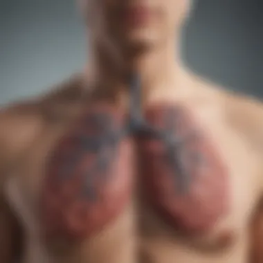
Visual Representation of Lung Cancer Insights


Intro
Lung cancer remains a prominent and complex health issue globally. Its visuals, from pathological slides to radiographic images, play a vital role in comprehending the disease. This section aims to impart comprehension of how imagery influences the understanding, diagnosis, and treatment of lung cancer.
Imagery serves as a powerful tool that bridges the gap between abstract scientific concepts and real-world implications for patients and healthcare providers alike. With the rise of advanced imaging technologies, there is an increasing need to analyze their impact on various stakeholders involved in the lung cancer spectrum.
Research Overview
This section outlines the methodologies that researchers employ to capture and interpret lung cancer imagery.
Methodological Approaches
In the realm of lung cancer research, scientists utilize several methodologies to acquire visual data. These include:
- Radiographic Imaging: Techniques such as X-rays, CT scans, and MRIs provide critical insights into the presence and stage of lung cancer.
- Pathological Assessment: Visualizations derived from biopsy samples are crucial for determining the type of cancer and its aggressiveness.
- Digital Modeling: Emerging technologies allow for simulation and 3D modeling of lung tumors, which offers new angles for analysis.
Utilizing these methods gives medical professionals a comprehensive view of the disease and its progression.
Significance and Implications
The significance of visual representations in lung cancer cannot be overstated. They serve several vital purposes:
- Enhanced Diagnostic Accuracy: Radiographic images lead to more precise diagnosis, improving patient outcomes.
- Informed Treatment Plans: Clear visuals enable oncologists to design personalized treatment regimens.
- Education and Awareness: Effective imagery can help inform patients and the public about lung cancer, consequently raising awareness.
"Visual aids in medical diagnostics bridge the gap between information and understanding, thus enhancing patient care."
Current Trends in Science
Imaging science continues to evolve, bringing innovative techniques and methodologies to the forefront of lung cancer research.
Innovative Techniques and Tools
Recent advancements include:
- AI and Machine Learning: These technologies are being integrated into image analysis to identify patterns that may go unnoticed by the human eye.
- Augmented Reality (AR): AR tools are assisting surgeons in visualizing tumors during operations, leading to improved precision.
Interdisciplinary Connections
The role of visual representation extends beyond oncology. Researchers from various fields collaborate to maximize the impact of imaging. For instance:
- Collaboration between Radiologists and Pathologists: Joint efforts enhance diagnostic accuracy through comprehensive assessments of visuals.
- Engagement with Public Health Experts: Shared knowledge improves community outreach and education regarding lung cancer.
Prologue to Lung Cancer
Lung cancer remains one of the most pressing health issues globally. Its significance in medical research and public health conversations cannot be overstated. Understanding lung cancer is critical for several reasons, notably its prevalence, mortality rates, and the challenges involved in early detection. This section lays the groundwork for a more comprehensive exploration of the visual representations relevant to lung cancer and the vital role they play in various processes, from diagnosis to education.
The complex nature of lung cancer involves not only its biological mechanisms but also its socio-economic impacts. By contextualizing lung cancer within a broader narrative, we can appreciate the implications for individuals and society as a whole. Additionally, the role of imagery in this field cannot be ignored. It serves as a bridge, facilitating better understanding among patients, healthcare providers, and the general public.
Definitions and Types
Lung cancer primarily refers to the uncontrolled growth of abnormal cells in one or both lungs. These cells can form tumors that may impede lung function, resulting in severe health complications. There are mainly two types of lung cancer: non-small cell lung cancer (NSCLC) and small cell lung cancer (SCLC).
- Non-Small Cell Lung Cancer (NSCLC) is the most common form, accounting for about 85% of all cases. It is further classified into several subtypes, including adenocarcinoma, squamous cell carcinoma, and large cell carcinoma.
- Small Cell Lung Cancer (SCLC), though less common, is more aggressive and tends to spread rapidly.
Each type exhibits distinct biological behaviors and responses to treatment, making their identification crucial for effective management.
Epidemiology and Statistics
Epidemiological data highlights the alarming incidence of lung cancer worldwide. According to the World Health Organization, lung cancer is the leading cause of cancer-related deaths. In 2020, approximately 1.8 million deaths were attributed to this disease. The statistics reveal stark differences in prevalence based on various factors such as geography, age, gender, and smoking status.
Some notable statistics include:
- Approximately 80% of lung cancer cases are caused by smoking.
- The five-year survival rate for lung cancer remains low compared to other cancers, at around 19%.
- Rates of lung cancer have been decreasing in some regions due to successful anti-smoking campaigns, yet the incidence remains high in others.
This data underscores the importance of targeted public health strategies and ongoing research in both epidemiology and imaging technologies.
Importance of Images in Medical Science
Images play a critical role in the field of medical science. Their ability to convey complex information quickly and effectively makes them indispensable in various medical specialties, especially in the diagnosis and treatment of diseases like lung cancer. This segment will underscore the significance of visual elements in enhancing understanding and facilitating communication among healthcare providers, patients, and researchers.
The integration of images within clinical settings transforms traditional data into visually accessible formats. They offer valuable insights without the need for extensive technical language, allowing for clearer interpretation of medical findings. For instance, radiographic images, which include X-rays and CT scans, enable clinicians to observe internal structures, identify abnormalities, and assess disease progression. This visual aid enhances the diagnostic process, making it more efficient and precise.
Additionally, images provide a universal language for communication among healthcare professionals. Regardless of their backgrounds, doctors, nurses, and specialists can interpret visual data, fostering collaboration and improving patient outcomes through shared understanding.
Key Benefits of Using Images in Medical Science:
- Improved Diagnosis: Higher accuracy in identifying conditions leads to timely interventions.
- Enhanced Patient Communication: Visual representation aids in explaining conditions, treatment options, and prognoses, enhancing patient understanding.
- Research and Education: Images are vital in academic settings for teaching purposes and disseminating research findings, making them essential tools in both learning and knowledge expansion.
Role of Visuals in Diagnosis
The role of visuals in diagnosis cannot be understated. Medical imaging techniques such as X-rays, CT scans, and MRIs are fundamental to establishing accurate diagnoses. They provide visual representations of the internal anatomy, revealing tumors, lesions, and other anomalies that may not be apparent through physical examinations alone.


Visuals allow for immediate assessment and monitoring of changes in the patient’s condition over time. For instance, a radiologist reviewing a chest X-ray can swiftly identify the presence of lung cancer and characterize it based on size, location, and density. This information is pivotal for determining the appropriate course of action.
Moreover, visual tools facilitate quicker decisions in emergency situations. The ability to acquire and analyze images rapidly can significantly impact patient survival rates, particularly in acute cases where lung cancer symptoms may overlap with other urgent medical conditions.
Visuals for Patient Understanding
In the context of patient education, visuals serve as a bridge between complex medical concepts and patient comprehension. They help demystify medical conditions for patients, offering clarity about lung cancer diagnosis and treatment options. For many patients, this understanding alleviates anxiety and encourages active participation in their healthcare decisions.
Examples of Visual Aids for Patients:
- Infographics: These can summarize treatment plans or explain the progression of lung cancer in simple terms.
- Illustrative Diagrams: Visual representations of healthy versus cancerous lung tissue help patients grasp the seriousness of their condition.
Furthermore, visuals enable informed discussions between healthcare providers and patients. When doctors can visually depict a tumor’s size and location on a scan, patients can better understand the implications for their treatment.
"Visual aids can profoundly influence a patient's grasp of their medical condition, fostering empowerment through knowledge."
In summary, the role of visuals in diagnosis and patient understanding is vital in the medical field, particularly concerning lung cancer. Across different stakeholders, these images enhance communication, improve educational efforts, and support better patient outcomes.
Radiographic Images of Lung Cancer
Radiographic images play a crucial role in the diagnosis and management of lung cancer. They allow for the visualization of internal structures, helping healthcare professionals understand the state of lung health. This section dives into specific types of radiographic images used most commonly: chest X-rays and CT scans. Each has its unique advantages and applications. Understanding these imaging modalities can significantly improve early detection and treatment planning for lung cancer.
Chest X-rays
Chest X-rays are often the first imaging test performed when lung cancer is suspected. They can reveal abnormalities in the lungs such as tumor presence, pleural effusion, and possible metastasis. The procedure itself is quick and relatively simple, making it widely accessible. However, it is important to note that the sensitivity of chest X-rays is limited compared to more advanced imaging techniques.
Some relevant facts about chest X-rays include:
- Easy and fast to perform.
- Affordable, making it a widely utilized screening tool.
- Capable of providing a basic overview of lung structures.
- Limited ability to detect small tumors or early-stage cancers; abnormalities can often appear similar to benign conditions.
Despite these limitations, chest X-rays serve an essential purpose in the initial evaluation of lung cancer. They can guide further imaging investigations and help in assessing the overall severity of any detected abnormalities.
CT Scans
Computed Tomography (CT) scans offer a more detailed view of the lungs compared to chest X-rays. This imaging modality uses a series of X-rays taken from different angles and compiles them to create cross-sectional images of the lungs. This level of detail helps in identifying small tumors that might not be visible on standard X-rays.
Advantages of CT scans include:
- High sensitivity to detect lung nodules and masses.
- Ability to assess tumor size and location precisely.
- Facilitation of planning for surgical interventions and radiation therapy.
- Potential use in monitoring treatment responses over time.
CT scans can also aid in staging lung cancer, which is vital for treatment decisions. Evaluating the extent of disease involvement is essential for determining prognosis and selecting the best therapeutic strategies.
Overall, both chest X-rays and CT scans form the backbone of radiographic assessment in lung cancer. Their unique strengths complement each other and contribute greatly to timely and effective patient care.
Pathological Images
Pathological images play a critical role in lung cancer diagnostics and research. They help in the assessment of tumor characteristics at the cellular level. The significance of these images cannot be understated as they offer insights into the biological behavior of cancer. By closely examining tissue samples, pathologists can provide essential information that guides clinical decisions. This analysis serves not just for diagnostic purposes, but also for understanding tumor progression, which is vital for treatment planning.
Biopsy Samples
Biopsy samples are among the most valuable pathological images in lung cancer diagnosis. A biopsy involves the removal of a small amount of tissue, which is then examined under a microscope. The importance of biopsy samples lies in their ability to confirm the presence of cancer. There are different types of biopsy methods, including fine needle aspiration, core needle biopsy, and surgical biopsy. Each method has its indications based on the tumor's location and size.
Key benefits of analyzing biopsy samples include:
- Tumor Type Identification: Accurate differentiation between types of lung cancer, such as adenocarcinoma or squamous cell carcinoma.
- Molecular Characterization: Detailed insights into genetic mutations, informing targeted therapies.
- Grading and Staging: Understanding the aggressiveness of the tumor helps in clinical decision-making.
Tissue Microarrays
Tissue microarrays offer a high-throughput method for analyzing multiple biopsy samples simultaneously. This technology enables researchers to study a variety of tissue samples in a single slide. The capability to compare several samples at once greatly enhances the efficiency of research into lung cancer.
The advantages of using tissue microarrays include:
- Standardization: Facilitates the comparison of different samples under controlled conditions.
- Reduced Costs: More samples can be analyzed at a lower cost than traditional methods.
- High Throughput: Allows for large-scale studies, which is essential for developing new treatment strategies.
"Pathological images not only confirm the diagnosis but also shape the treatment strategy for lung cancer patients."
Comparative Images
The use of comparative images is critical in the evaluation and understanding of lung cancer. By providing visual contrasts between normal and cancerous lung tissue, these images serve as a powerful educational tool. They highlight the physical differences that are often subtle yet impactful. Comparative images can facilitate better comprehension of lung cancer's progression and variations, which is essential for both medical professionals and patients alike.
In essence, comparative imagery extracts complexity and presents it in a more digestible format. This visual approach can aid in diagnostic accuracy, allowing radiologists and oncologists to discern malignancies more precisely. Furthermore, these images can assist students and researchers in visualizing the pathology of lung cancer, enabling a deeper understanding of disease mechanisms.
Normal Lung vs. Cancerous Lung
Comparative images of normal lung versus cancerous lung tissue reveal stark differences in structure and appearance. Normal lung tissue appears uniform and healthy, characterized by its spongy texture and a network of airways visible through imaging. In contrast, cancerous lung tissue often displays irregularities. This might include masses, nodules, or areas of consolidation that signal tumor presence.
These visual distinctions do not just assist in diagnosis; they also convey the severity and stage of the disease. For instance, early-stage lung cancer may show localized lesions, whereas late-stage disease may present with widespread metastasis. By examining these images closely, medical personnel can formulate more accurate treatment plans tailored to the specific type and stage of lung cancer.
Comparison of Different Lung Cancer Types
Lung cancer is not a monolithic disease; it encompasses various types, such as non-small cell lung cancer and small cell lung cancer. Comparative images can elucidate the phenotypic distinctions between these types, aiding in both diagnosis and treatment strategies.
For example, non-small cell lung cancer tends to manifest as larger, more well-defined masses on imaging studies, while small cell lung cancer often presents with more diffuse infiltrative patterns. Understanding these differences is paramount for oncologists as they guide treatment.


- Non-small cell lung cancer (NSCLC): Common format includes adenocarcinoma, squamous cell carcinoma, and large cell carcinoma. Imaging shows larger, irregular masses.
- Small cell lung cancer (SCLC): Characterized by smaller, more invasive tumors that may lead to early metastissues.
Advancements in Imaging Technology
The field of lung cancer diagnosis and treatment is constantly evolving, thanks to significant advancements in imaging technology. These innovations are essential as they enhance the accuracy of detection and monitoring throughout the disease's progression. The integration of new imaging modalities not only improves clinical outcomes but also augments the overall understanding of lung cancer among both healthcare professionals and patients. The use of advanced imaging techniques leads to better stratification of therapy, fostering personalized treatment plans tailored to individual patient needs.
3D Imaging Techniques
Three-dimensional imaging techniques provide a more comprehensive view of lung anatomy and pathology compared to traditional two-dimensional methods. These 3D images allow for detailed visualization of tumors, facilitating precise measurements and accurate assessments of their characteristics. Computed tomography (CT) scans have evolved from simple flat images to sophisticated 3D models that can be manipulated for enhanced visualization.
- Benefits of 3D imaging include:
- Improved tumor localization
- Enhanced surgical planning
- Better imaging for radiotherapy
The 3D reconstruction offers oncologists multidimensional perspectives that inform decision-making during treatment planning. For instance, surgeons can visualize the relationship between a tumor and surrounding tissues or structures, which is crucial for achieving optimal surgical outcomes. This type of imaging minimizes unnecessary invasiveness while maximizing treatment effectiveness.
Artificial Intelligence in Imaging
Artificial intelligence (AI) is becoming increasingly integral in lung cancer imaging. AI algorithms can analyze vast amounts of imaging data with remarkable speed and accuracy, providing support in identifying potential malignancies that may be overlooked by the human eye. This capability is particularly important in early-stage lung cancer detection, which can significantly affect prognosis and survival rates.
- Key contributions of AI in imaging include:
- Automated detection of anomalies
- Enhanced image processing
- Predictive analytics for treatment responsiveness
AI tools are capable of learning from existing imaging data, continuously improving their predictive abilities. Furthermore, as imaging databases grow, AI systems can assist radiologists by highlighting areas of concern, thus streamlining the interpretation process. This collaboration between technology and medical professionals is vital for enhancing diagnostic accuracy and ensuring that patients receive timely interventions. > "AI's role in imaging represents a shift towards precision healthcare, allowing for personalized treatment approaches that are informed by data-driven insights."
In summary, advancements in imaging technology, particularly through the implementation of 3D imaging techniques and artificial intelligence, offers remarkable benefits in the landscape of lung cancer diagnosis and treatment. These innovations promise not only to improve patient outcomes but also to enrich the knowledge and skills of healthcare providers.
Image Interpretation and Analysis
Image interpretation for lung cancer is a multifaceted process that combines various skills and expertise. Understanding the nuances in imaging can significantly influence diagnostic accuracy and treatment options. It extends beyond just viewing data; it encompasses critical analysis of various imaging modalities, including radiographic and pathological visuals. Proper interpretation is essential as it serves as a precursor to understanding the disease's progression, treatment choices, and patient outcomes.
Images can reveal cellular changes indicative of lung cancer, which may not be visible through other diagnostic methods. Interpreting these changes involves recognizing patterns and anomalies that could signify malignancy. Moreover, the interpretation must also consider a host of factors, including patient history, clinical findings, and other diagnostic results.
Challenges in Image Assessment
Assessing lung cancer images poses several challenges. One significant obstacle is the variability in image quality. Factors such as equipment used, settings, and the patient's anatomy can create inconsistencies. High-quality images are crucial for accurate diagnosis, and poor-quality images can lead to misinterpretation.
Another issue lies in the subjective nature of image analysis. Different radiologists and oncologists may arrive at divergent conclusions based on the same set of images. This discrepancy can stem from personal experience, the interpretative framework used, and familiarity with lung cancer imaging. To mitigate this, standardized protocols and training programs are essential.
Additionally, certain imaging features may overlap with benign conditions, complicating the assessment process. Distinguishing between potentially malignant and harmless findings is critical yet often complicated.
Key Challenges Summary:
- Variability in image quality
- Subjective interpretations by different specialists
- Overlapping features with benign conditions
Role of Oncologists and Radiologists
Oncologists and radiologists play pivotal roles in the image interpretation process. Radiologists are trained to analyze images and derive preliminary conclusions about the presence of lung cancer. Their expertise encompasses understanding the subtleties in imaging that might elude other specialists.
Oncologists complement this by integrating imaging findings with clinical data. They bring in the clinical context, focusing on how imaging correlates with symptoms and laboratory results. This collaboration is vital for forming an accurate diagnosis and tailored treatment strategies.
Moreover, communication between radiologists and oncologists improves patient outcomes. When both specialists engage in discussions about imaging findings, it fosters a better understanding of the disease. Their combined knowledge enhances decision-making concerning further investigations and treatment plans.
“Interpretation of imaging is not just about the images; it also involves patient context and expert collaboration.”
Maintaining a multidisciplinary approach is crucial since it ensures that all relevant perspectives are integrated into the care plan. Continuous education and dialogue between these professionals remain paramount for ongoing improvement in imaging techniques and interpretation practices.
Patient Education Through Images
Patient education regarding lung cancer is crucial, and images play a significant role in this process. Visual representation serves multiple functions, including simplifying complex medical concepts and enhancing understanding among patients. In a field where technical jargon and intricate anatomical details can overwhelm individuals, images act as a bridge to facilitate comprehension.
Visual tools provide clarity, particularly when explaining diagnostic procedures, treatment options, and the nature of the disease itself. For instance, an annotated image of lung anatomy can help patients identify where tumors might develop, while visual guides portraying the stages of cancer progression can demystify their journey. Such clarity is essential for informed decision-making and fosters an environment where patients feel more engaged in their care.
Moreover, visual aids can alleviate anxiety associated with medical consultations. They offer a way for patients to visualize their condition and the path forward, which can be comforting in stressful times. This approach enhances the overall experience of learning about lung cancer, making it a critical component of patient education.
Visual Guides for Patients
Visual guides play a pivotal role in helping lung cancer patients navigate their diagnosis and treatment options. These guides typically include diagrams, charts, and infographics designed to present information in an easily digestible format. Patients often struggle to understand medical terminology, but visual representations break down these barriers.
Images showing different types of lung cancer can provide clarity about the various forms of the disease, such as non-small cell lung cancer and small cell lung cancer. Additionally, visual timelines can illustrate the treatment process, highlighting milestones such as chemotherapy cycles or surgery. This visual context enhances a patient’s appreciation for what to expect during their journey.
Furthermore, accompanying images with explanatory text can reinforce learning. For instance, a visual guide on potential side effects of treatment can prepare patients for what they might experience, thus making them feel more in control.
"Visual tools not only aid understanding but also empower patients to become active participants in their healthcare decisions."
Informed Decision-Making
Informed decision-making is a cornerstone of patient autonomy, and visual aids significantly contribute to this process. When patients have access to relevant images, they can better assess their options in consultation with healthcare professionals. This is particularly vital in the context of lung cancer, where treatment decisions can be complex and varied.
Images that depict various treatment modalities, including surgical options and targeted therapies, allow patients to visualize their choices. Understanding the differences between treatments helps patients weigh the benefits and risks involved, leading to more personalized and appropriate decisions.
Moreover, engaging with visual content fosters discussions between patients and their medical teams. When patients can physically see representations of treatment outcomes or potential complications, they are more likely to ask informed questions and express their concerns. This two-way communication is essential for fostering a trusting relationship between patients and healthcare providers.


In summary, incorporating images into patient education enhances understanding and supports informed decision-making. Visual tools serve as an effective strategy to empower patients, making them feel more comfortable and confident in navigating their lung cancer journey.
Public Awareness and Campaigns
Public awareness and campaigns about lung cancer play a crucial role in enhancing knowledge regarding this disease. Effective communication strategies can bridge the gap between complex medical information and general understanding, which is vital for early detection and treatment. Campaigning is not just about spreading awareness; it involves educating people about the risks, symptoms, and available treatments. This is especially important given the significant stigma attached to lung cancer, often perceived as a "smoker's disease". Changing public perception can encourage those at risk to seek medical advice.
Using Images for Advocacy
Images serve as powerful tools for advocacy in lung cancer campaigns. A well-placed photograph showing the impact of the disease can convey urgency and empathy in ways that words alone cannot. Here are some ways that imagery can be utilized:
- Illustrative Impact: Graphic images of diseased lungs can highlight the severity of lung cancer, resonating emotionally with individuals who may not be aware of how dangerous this disease can be.
- Informational Graphics: Infographics displaying statistics on lung cancer can effectively communicate essential data, such as survival rates or risk factors, making it easier for the audience to digest information quickly.
- Storytelling through Visuals: Images of survivors or patients can humanize the disease, making it relatable. Personal stories complemented by photographs can motivate viewers to participate in lung cancer awareness events.
By leveraging images strategically, advocates can thrust the issue of lung cancer into the forefront of public discourse, pushing for better funding, research, and support systems.
Social Media and Visual Storytelling
Social media platforms have transformed how information is shared and consumed. Through visual storytelling, organizations and advocates can create engaging narratives around lung cancer that foster understanding and action. The immediacy of social media allows campaigns to reach diverse audiences quickly. Some effective strategies include:
- Shareable Content: Create visually appealing posts that are easy to share. Memes or short videos discussing lung cancer facts can go viral, expanding reach.
- Engagement through Visual Challenges: Campaigns can host challenges that encourage users to share personal lung cancer stories or awareness images, prompting community interaction.
- Live Events with Visual Components: Hosting webinars or live Q&A sessions with oncologists, using visuals to explain treatment options or how to conduct self-examinations, gives patients direct access to information.
In the realm of social media, visuals are not merely supplementary; they are essential. A post with a well-crafted image is more likely to attract attention than text alone.
Understanding lung cancer and its implications will take a collective effort, led by informed and engaged communities through effective visual storytelling.
Future Directions in Imaging for Lung Cancer
As we explore the realm of lung cancer imaging, it is essential to consider the future directions in this field. Advancements in technology signify significant potential in improving diagnostic accuracy and treatment pathways for lung cancer patients. The ability to visualize the disease in more sophisticated ways not only enhances the understanding but also aids in targeted therapies. This section delves into the emerging technologies that promise to reshape the landscape of lung cancer imaging, as well as the challenges that may arise during their implementation.
Emerging Technologies
Recent strides in imaging technology offer several promising solutions for lung cancer diagnosis and management. Key innovations include:
- High-Resolution Imaging: Advanced imaging methods, such as ultra-high-resolution CT scans, allow for more precise visualization of small lung nodules, potentially leading to earlier detection of cancerous changes.
- Positron Emission Tomography (PET): This technique, especially when used in combination with CT scans, provides metabolic information about lung lesions, helping to differentiate between benign and malignant growths.
- Radiomics: This emerging field focuses on extracting large amounts of quantitative features from medical images using data-characterization algorithms. Radiomics could lead to more personalized treatment plans based on the specific characteristics of both the tumor and the patient.
- Machine Learning: The application of artificial intelligence in analyzing imaging data enhances pattern recognition, assisting radiologists in detecting minute discrepancies in lung images that human eyes might miss.
These technologies pave the way for a more detailed assessment of lung cancer, improving patient outcomes. They enable clinicians to tailor treatments based on a more comprehensive understanding of each patient's condition. However, alongside these advancements come substantial challenges.
Challenges Ahead in Implementation
The path to integrating novel imaging technologies into standard clinical practice is fraught with challenges. Some of these include:
- Cost and Accessibility: High-end imaging technologies often require significant investment. Many healthcare facilities may lack the resources to acquire and maintain such equipment, leading to disparities in access to cutting-edge imaging services.
- Training and Expertise: The successful implementation of emerging technologies depends heavily on the training of medical personnel. Radiologists and technicians must be proficient in new methodologies to maximize their utility and ensure accurate interpretations.
- Data Integration: Merging data from various imaging modalities and ensuring that different systems interface effectively can be technically complex. A seamless data integration process is essential for effective decision-making.
- Regulatory and Ethical Issues: As technologies advance, they also raise questions regarding privacy and data security, especially concerning patient information. Adhering to regulations is crucial yet poses another layer of complexity.
In summary, while the future directions in imaging for lung cancer present an array of opportunities to enhance diagnosis and treatment, careful consideration must be given to the accompanying challenges. Stakeholders must remain vigilant and proactive to ensure these advancements translate into real-world benefits for lung cancer patients.
Ethical Considerations in Imaging
The visual representation of lung cancer is not just about the images themselves; ethical considerations play a crucial role in how these visuals are created, shared, and utilized. Ethics in medical imaging encompass various dimensions, including patient privacy, data security, and the informed consent process. Adhering to ethical guidelines ensures that the use of images contributes positively to patient education and medical research while respecting individual rights and societal norms.
Patient Privacy and Data Security
Patient privacy is a fundamental concern in the medical field, especially in imaging, where sensitive data is often involved. Radiographic and pathological images contain identifiable information that can link them back to specific individuals. Maintaining the confidentiality of this information is essential. Medical institutions must implement robust data protection protocols to prevent unauthorized access and breaches.
In recent years, several high-profile data breaches have highlighted the risks associated with storing medical images. These incidents not only compromise patient trust but can also lead to significant legal and financial repercussions for healthcare institutions. Effective strategies for protecting patient data include:
- Encryption of images: Using encryption technology helps to secure images during transmission and storage.
- Access controls: Ensuring that only authorized personnel can access sensitive data.
- Regular audits: Conducting routine assessments of data security measures to identify and rectify vulnerabilities.
"A patient’s right to privacy in their medical data is as critical as the care they receive."
Consent for Image Use
Informed consent is a pivotal component of ethical imaging practices. Patients must be adequately informed about how their images will be used. This includes details on whether their images will be utilized for educational purposes, research, or public awareness campaigns. The consent process should be clear and transparent, allowing patients to make informed decisions about their participation.
Acquiring consent is not just a regulatory requirement; it also fosters trust between patients and healthcare professionals. To ensure an ethical approach to consent, medical institutions should consider the following:
- Clear communication: Use simple language to explain how images will be used and the potential risks involved.
- Voluntary participation: Ensure patients know they can decline without affecting their care.
- Reassurance of anonymity: Explain how patient identity will be protected if their images are used publicly.
The ethical management of patient privacy and consent is not merely a box to check; it is a commitment to uphold the dignity and autonomy of patients. By establishing strong ethical guidelines, the medical community can enhance the integrity of their practices in imaging while advancing knowledge and awareness of lung cancer.
Epilogue
The conclusions drawn in this article underscore the significant role of visual representation in the understanding and management of lung cancer. The visual tools discussed amplify the capability of healthcare professionals to detect, diagnose, and treat this complex disease effectively. Accurate imaging methods, such as radiographic and pathological images, along with emerging technologies, facilitate a clearer understanding of the tumor's characteristics and behavior.
Additionally, visuals serve as an essential bridge between the medical community and patients. They promote health literacy, ensuring that individuals grasp their condition better and enhancing informed decision-making. Images enable patients to visualize the process of the disease, which can alleviate some anxiety associated with diagnosis and treatment options.
In addressing public awareness, visual representation acts as a powerful advocacy tool. Campaigns utilizing images effectively communicate the risks and realities of lung cancer, engaging a broader audience to foster better understanding and support for research initiatives.
Overall, the convergence of imaging technology and visual representation in the context of lung cancer is vital. It not only enriches clinical practice but also contributes to a comprehensive understanding of the disease across various stakeholders.
Recap of Key Points
- Role of Images in Medical Science: Images provide crucial support in diagnosis and enhance communication between healthcare providers and patients.
- Advancements in Imaging Technology: Innovations like AI and 3D imaging facilitate better detection and monitoring of lung cancer.
- Importance for Patient Education: Visuals assist patients in grasping their diagnosis and treatment options, leading to more active participation in their healthcare.
- Public Awareness Campaigns: Images are pivotal in advocacy efforts, raising awareness about lung cancer and promoting research funding.
Final Thoughts on Lung Cancer Imaging
The landscape of lung cancer management is continually evolving, with imaging technology at the forefront of these advancements. The benefits of using visual representation extend far beyond clinical settings— they reach into patient education and public health advocacy. As technology progresses, the potential for more effective imaging solutions will likely enhance our understanding and treatment of lung cancer.
Investing in proper training for healthcare providers to interpret these images accurately and communicating findings effectively with patients remains essential. Additionally, ethical considerations, including patient privacy and consent, must continue to guide the integration of imaging in lung cancer management.
In summary, the future of lung cancer imaging is promising, holding the potential to transform patient outcomes significantly. The emphasis on visual representation will remain critical in ensuring all stakeholders— from researchers to patients— are well-informed and engaged in the fight against lung cancer.


